Autologous therapy is changing how reconstructive surgeons plan grafts and manage donor sites, improving patient outcomes
By Corianne Rogers, MD
Reconstructive surgery has always been about more than closing a wound. The goal is to restore both form and function, providing durable coverage that protects, while achieving results that look and feel as natural as possible. Nowhere is this tested more than with acute injuries. In the United States, nearly 500,000 people are treated for burns each year, and roughly 40,000 require hospitalization.1 Beyond burns, there are traumatic wounds, closure complications, and defects after skin cancer resection that fill a reconstructive practice. A constant reminder to surgeons that covering the wound is only the starting point, and that success should be measured by how closely the rebuilt skin returns a patient to the way they looked and lived before.
Over the past decade, autologous skin cell therapy, commonly known as Spray-On Skin Cells, has become one of the tools that has meaningfully advanced that goal. The technology has changed not just how surgeons approach complex cases but also raised the bar for reconstruction techniques in ways that constantly surprise and excite us.
By working with the body’s own biology rather than around it, these therapies have expanded what is achievable with the tools reconstructive surgeons already use.
How It Works
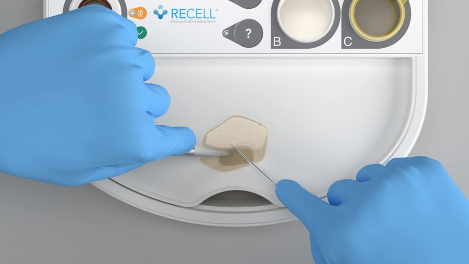
Using a specialized point-of-care device called RECELL, from Avita Medical, clinicians prepare a suspension of Spray-On Skin Cells from a sample of the patient’s skin, often the size of a postage stamp. The suspension contains several cell types, each with a defined role in rebuilding skin: keratinocytes regenerate the epidermis, papillary dermal fibroblasts contribute to the creation of new extracellular matrix proteins, and melanocytes produce melanin that restores natural pigmentation.2 Delivered together across the wound bed, these cells support healing and re-epithelialization throughout the wound rather than only at its margins.
Because the cells are the patient’s own, there is no risk of rejection and no wait for an outside lab to culture cells. Preparation and application happen in the operating room during the same procedure, rather than over days or weeks as a cultured approach would require, removing a source of delay from the reconstructive workflow.
Donor skin is a limited resource, and harvesting it creates a second, often large, wound that can scar, hurt, and be prone to infection, slowing recovery. In patients with extensive injuries and little healthy skin to spare, this can become the limiting factor in the reconstruction. Turning the sample into an autologous cell suspension solves that problem because the surgeon is no longer trying to stretch donor tissue to cover more surface area, but instead distributing living cells across a larger surface. A single sample can treat an area as large as 80 times its own size, and clinical data show closure with up to 97% less donor skin compared with conventional split-thickness grafting,3 and up to 60% fewer autografting procedures.4
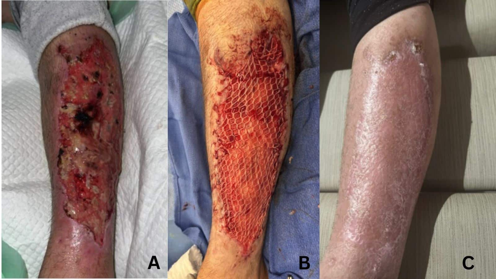
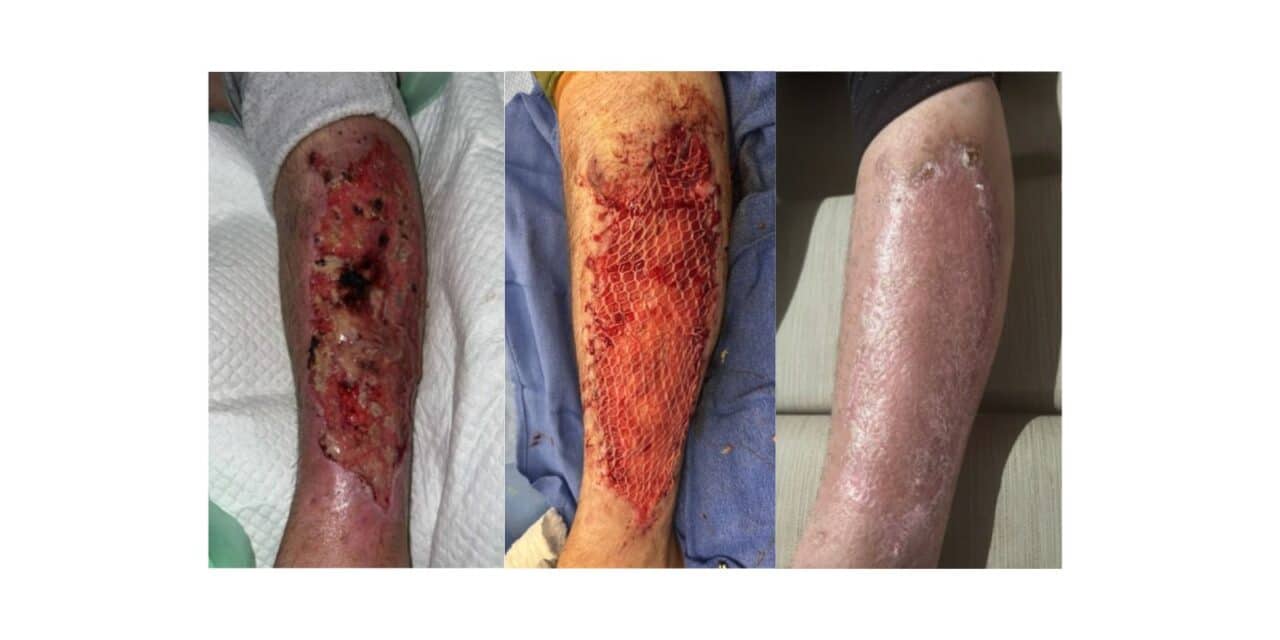
A 35-year-old female patient presented with 23 x 8.5 cm full-thickness left lower extremity wound following infection from IV drug use (Figure A). No comorbidities. Prior treatment: surgical debridement, piscine xenograft, compression dressing. A 3:1 mSTSG + RECELL application was performed (Figure B). Figure C shows would 2 months post-op.
Where Cell-Based Therapy Fits In Practice
Three use cases stand out where this technology consistently adds value.
The most straightforward is with large total body surface area burns, where harvestable skin is limited. Widely meshed grafts close faster and flatter, with better cosmetic results, when autologous cells are applied. The diamond-patterned appearance left by meshed grafting is one of the things that most troubles patients about how they look after recovery. When Spray-On Skin Cells are part of the plan, the pattern is meaningfully reduced, if visible at all.
The second is when flap and graft tissue meet. Known as the transition zone, it is often the most visually conspicuous part of the result and the area patients notice and ask about most. Regenerating the surrounding skin with the patient’s own cells can improve how those interfaces integrate and blend.
The third is the donor site itself. Spray-On Skin Cells can be used on the harvested site(s), providing faster, more comfortable, and improved cosmetic healing in the area that patients often report as the worst part of recovery.
In oncologic reconstruction, the same approach can be used after skin cancer resection. When combined with a dermal matrix in staged reconstructions of deeper defects, the cell-based suspension provides a reliable means of rebuilding a vascularized base over one to several weeks. Closing the surface occurs once the bed is ready. In a symbiotic relationship, the matrix provides the structural depth that the suspension cannot, and the cells supply the durable, pigmented epithelium that the matrix cannot generate on its own.
Grafts, flaps, and microsurgery will always have a place in reconstructive surgery. Cell-based therapies, however, are changing how surgeons use those tools and what they can achieve—not by replacing them, but by amplifying what they deliver.
Making the Operational Case
The clinical case is compelling, but the operational one is equally relevant to a practice. Preparing the suspension intraoperatively allows most closures to be performed in a single trip to the OR rather than a staged series, freeing operative time and beds.
Real-world registry data from U.S. burn centers show that patients with deep partial-thickness burns treated with autologous skin cell suspension experienced 36% shorter hospital stays, or roughly 5.6 days, compared with traditional skin grafting, translating to an estimated $42,000 in reduced care costs per case and the capacity to treat approximately 13 additional patients per bed annually.5 Health-economic analyses further support the downstream benefits, with demonstrated reductions in autografting procedures and overall per-case costs.6
For surgeons making the case to their institutions, the data translate directly into language that hospital administrators respond to: shorter stays, lower costs, and better bed utilization. In a resource-constrained environment, a technology that reduces procedural complexity while improving outcomes is a relatively straightforward clinical and financial argument.
The Future of Dermal Regeneration
Re-epithelialization is where cell therapy works today. The future is headed toward dermal regeneration. The xenograft options currently available for dermal substitution are not always successful, and they often lack the elasticity and resilience of native skin. A more reliable way to regenerate the dermis would allow surgeons to restore not only the surface but also the dermis itself, along with appendages such as sweat glands and hair follicles, improving aesthetics and function, while reducing contracture rates, restoring natural elasticity, and lowering the risk of scarring. For patients, that would mean reconstructions that move and feel more like their original skin.
While that capability is still being explored, the progress already achieved with autologous cell therapies has expanded what reconstructive surgeons can accomplish today. As clinical experience and evidence continue to grow, these therapies are helping move reconstruction closer to its ultimate goal of restoring skin that looks, feels, and functions as naturally as possible. PSP
Clinical images courtesy of Dr Corianne Rogers. Recell image courtesy of AVITA Medical.
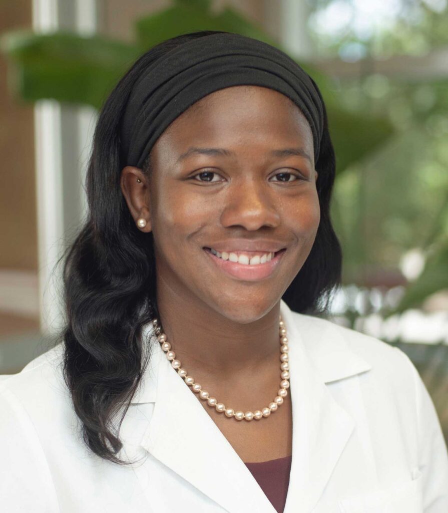
Corianne Rogers, MD, is a board-certified plastic surgeon with fellowship training in hand surgery. She practices at WakeMed Health & Hospitals in her hometown of Raleigh, NC. Her practice is a mixture of general plastic & reconstructive surgery, including trauma, burn, chronic wounds, and oncologic reconstruction, with a focus on lower extremity salvage and hand surgery.
References
- National Hospital Ambulatory Medical Care Survey: 2021. Centers for Disease Control and Prevention. https://www.cdc.gov/nchs/data/nhamcs/web_tables/2021-nhamcs-edweb-tables-508.pdf. Accessed September 11, 2024.
- Wood FM, Giles N, Stevenson A, Rea S, Fear M. Characterisation of the cell suspension harvested from the dermal epidermal junction using a ReCell® kit. Burns. 2012;38(5):723-729. doi:10.1016/j.burns.2011.03.001.
- Holmes JH 4th, Molnar JA, Shupp JW, et al. Demonstration of the safety and effectiveness of the RECELL® System combined with split-thickness meshed autografts for the reduction of donor skin to treat mixed-depth burn injuries. Burns. 2019;45(4):772-782. doi:10.1016/j.burns.2018.11.002.
- Foster K, Amani A, Carter D, et al. Evaluating health economic outcomes of autologous skin cell suspension (ASCS) for definitive closure in US burn care using contemporary real-world burn center data. J Curr Med Res Opin. 2021;4(11):1042-1054. doi:10.52845/CMRO/2021/4-11-1.
- Hoppe A, Carter J, Phillips B, et al. 891 Skin Is In: A national analysis of skin cell suspension autograft in burn centers. J Burn Care Res. 2025;46(1):S322. doi:10.1093/jbcr/iraf019.422.
- Kahn SA, Carter JE, Wilde S, et al. Autologous skin cell suspension for full-thickness skin defect reconstruction: current evidence and health economic expectations. Adv Ther. 2024;41(3):891-900. doi:10.1007/s12325-023-02777-7.