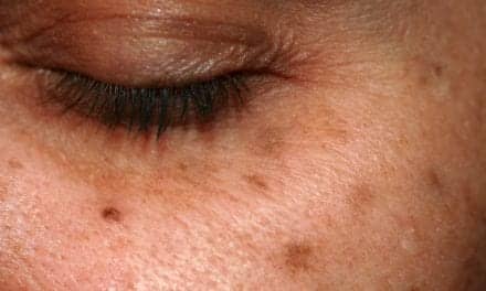
New imaging technology developed by engineers at the University of Washington may allow scientists to analyze what happens within the microvascular system during a “liquid facelift” and better manage complications.
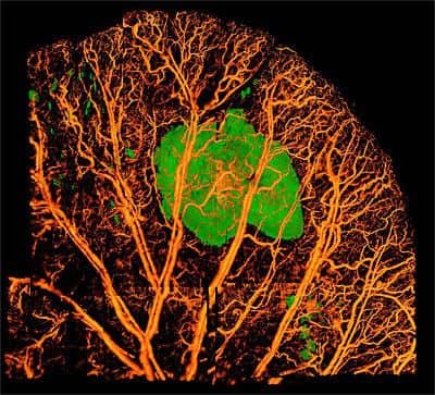
Using optical microangiography, researchers found that complications arose when filler was inadvertently injected into the bloodstream rather than in the intended soft tissues of the face. The gel builds up in a vessel, blocking blood movement and oxygen exchange. The team tested this in the ears of mice to show that filler injections into blood vessels are most likely the cause of tissue death and other complications associated with fillers (see image). Physicians can try to reverse the effect of vascular blockage with hyaluronidase.
The fine-resolution imaging produces 3-D images of the body’s vascular network by shining a light onto the tissue without touching it or adding any fluorescent dyes. It can visualize blood vessels as small as 5 microns in diameter.
The research team is now testing all types of available cosmetic fillers to see if their results hold on each brand and evaluating new treatments for reversing procedure complications.
Other potential applications of the technology include analyzing how wounds heal, tracking what happens during strokes and traumatic brain injuries, and imaging human eyes to study diseases such as glaucoma and macular degeneration.
Researchers presented the study results at the annual meeting of the American Society of Ophthalmic Plastic and Reconstructive Surgery.
Photo credit: Siavash Yousefi, U of Washington